
CTAB Extraction of Pocillopora meandrina
Low-Salt CTAB DNA Extraction Attempt for HMW DNA from Pocillopora meandrina for Genome Sequencing
Goal: Get 25ug of high quality/non-degraded HMW DNA from our Pocillopora meandrina sample
Results: Yield was not terribly low but not good enough, only ~13.5ug. Quality was poor, there is a lot of low base pair smearing in every sample
Major takeaways: This extraction yielded worse results than the genomic tip extraction, I can try again but it might not be worth it
Background
We are trying to extract HMW DNA from Pocillopora meandrina and Porites lobata samples to send for PacBio sequencing for genome building. These need to be great quality for long read sequencing, and have ~25ug total DNA. I have tired genomic tip extractions previously (x and x), however the sequencing company says they are too degraded and suggested we try a “CTAB protocol.” We have not used the CTAB (Cetyl trimethylammonium bromide) extraction protocol before, so I searched for one that seemed the most applicable to coral tissue, and found a modified low-salt CTAB procedure that works well for mollusk tissue, described here, with full protocol here. The DNA extracted in the paper looked like good quality (although only a gel was run), and the yield per extraction looked like it would be between 2-10ug. Yield of course can be dependent on the input tissue. I decided to do 6 extractions to hopefully get enough DNA yield. I followed the protocol almost exactly, however we did not have the same rotating incubation set up as in the protocol. Filter tips were used for all steps, and some steps used wide-bore tips (noted). Additionally, this protocol uses chloroform/isoamyalcohol which is very toxic and vaporous, all steps after the incubation are done in the hood except for centrifugation, solid and liquid hazardous waste containers were set up, and secondary containment of liquids was used. PPE of gloves, coat, mask, and glasses were worn. See below:
Buffer Prep
- CTAB Lysis Buffer 100mL
- 10mL 1M Tris HCL pH 8
- 4mL 0.5M EDTA
- 2g CTAB powder
- 7g NaCl molecular grade
- ~85mL ultra pure H2O (volume up to 100mL)
- Diluted CTAB Buffer (low salt) 100mL
- 10mL 1M Tris HCL pH 8
- 4mL 0.5M EDTA
- 2g CTAB powder
- ~85mL ultra pure H2O (volume up to 100mL)
- High Salt TE Buffer 50mL
- 2.907g molecular grade NaCl
- 200ul 0.5M EDTA
- 500ul 1M Tris HCl
- ultra pure water up to 50mL
- Note, I did not autoclave the TE buffer because the autoclave was broken, because we don’t autoclave most of our elution liquids I did not think this would be a problem, however it may have been
Sample Prep
- Set incubator genie to 60 degrees C
- Chilled mortar and pestle in -80
- Sterilized scraper with 10% bleach, DI, and 70% ETOH and placed in -80
- Prepared 6 DNA low bind tubes with 500ul each of CTAB lysis buffer
- Brought scale over to the bench
- Filled the mini dewar with LN2
- Took out a coral piece from the conical and weighed it on the scale inside the chilled mortar
- Weight: 1.81g
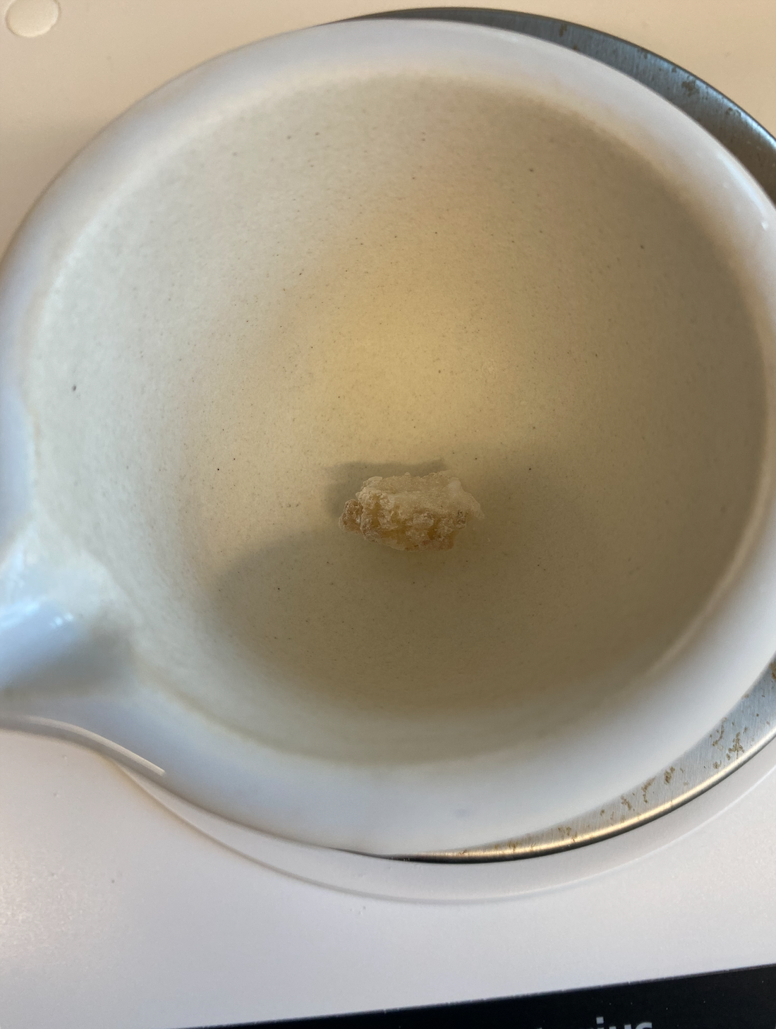
- Poured in LN2 multiple times and ground tissue until it was a powder, there was a lot of skeleton in there as well

- Used the chilled scraper to scrape some sample into each of the 6 1.5mL tubes with the CTAB buffer, not all the sample was used and the rest I scrapped back into the conical
- The tubes had a lot of sample in each
- I decided to add more CTAB buffer because I was worried I wouldn’t be able to get the full 500ul out in later steps because of all the skeleton, so I added 400ul more of CTAB lysis buffer to each tube, total of 900ul. Later this seemed like it may have been too much but at the time it was just a guess

- With the increased volume I had to increase the amount of Proteinase K proportionally, 30ul originally, now 54ul. I adde 54ul of Zymo Research proteinase K to each tube
- Tubes were vortexed and spun down, and placed in the incubator genie at 60 degrees C, rocking 25 speed, for 3 hours. The protocol suggests a rotating incubator at 150rpm, our thermomixers only have a lowest setting of 300rpm so I decided not to use them. I do not know how close 25 speed is on the rocking incubator to 150rpm, looking back it might have been too fast
DNA Separation and Precipitation
- After incubation, the sample liquid looked browner and the skeleton looked mostly white
- Transferred 500ul to new DNA low bind tubes using wide-bore pipette tips. There was some excess liquid in each tube that was not used
- Let liquid come to room temperature (5-7 minutes), and moved set up to the hood
- Added 500ul of chloroform/isoamyalcholol 24:1 to each tube and inverted 10 times to mix, the tubes became milky white
- Tubes were centrifuged for 15 minutes at room temp, 3,000 rcp (g)
- After centrifugation there was clear phase separation
- 400ul of the aqueous (top) layer was transferred to new DNA low bind tubes with wide bore tips
- Two volumes (800ul) of the diluted CTAB buffer were added to each tube and were inverted 5-10 times to mix
- The tubes were placed in the rocking incubator at 60 degrees C 25 speed for 30 minutes
- Here, crystals are supposed to form in the liquid, but “may be hard to see for some samples”. After 30 minutes, there were no visible crystals, although I am not sure what they are supposed to look like. The protocol says to increase incubation time and rotating speed. I incubated the tubes for another 30 minutes and increased the speed to the max, 35
- After 1 hour incubation I needed to stop to have time to finish the rest of the steps, there were still no crystals visible

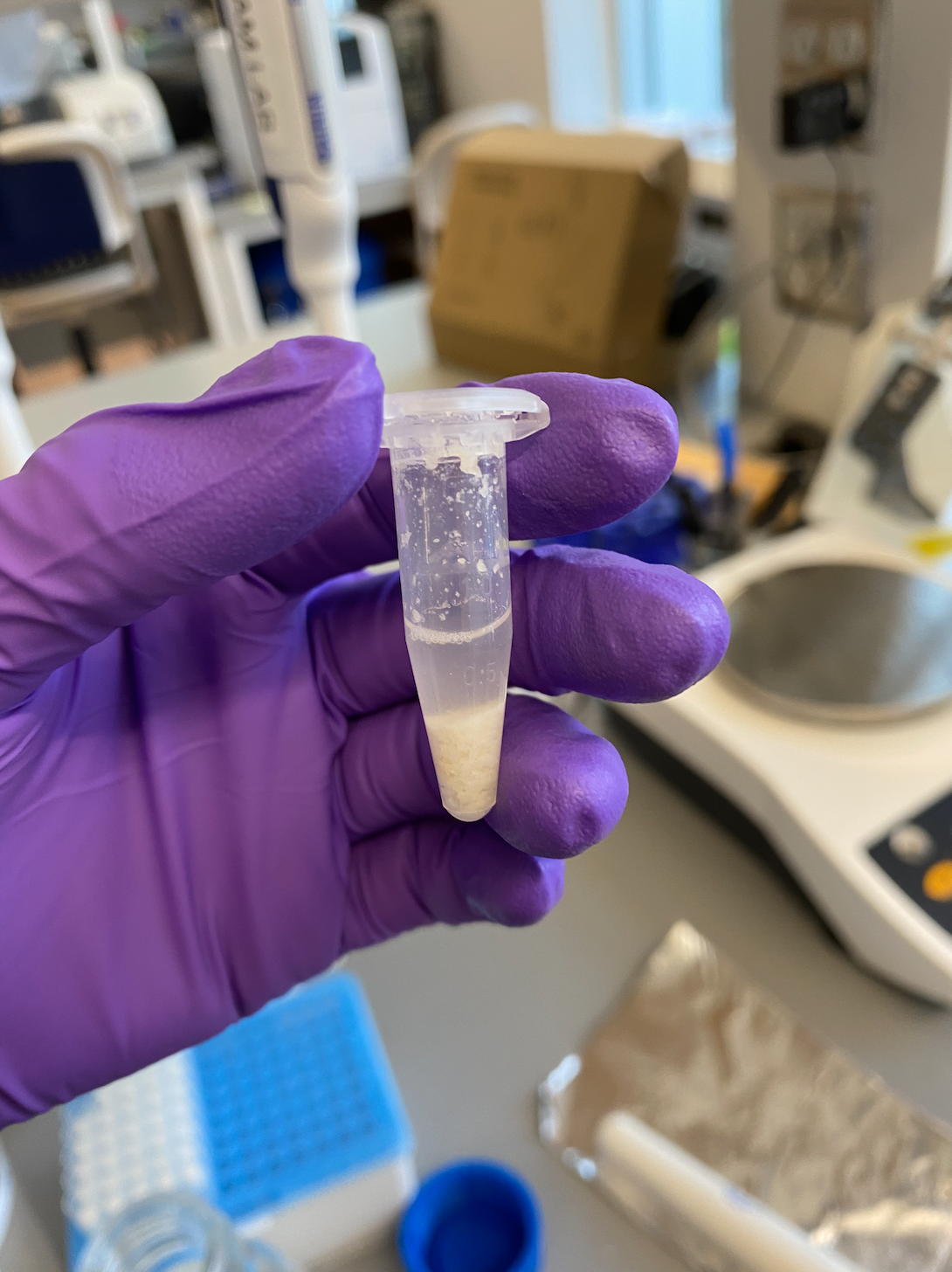
- The tubes were then centrifuged for 3 minutes at 16,000 rcf (g). There was a visible long pellet in each tube, so I guessed that crystals did form

- Removed and discarded the supernatant (hazardous)
- Added 1mL fresh 80% ethanol and inverted to mix, the pellet dissolved it seemed like
- Tubes soaked at room temp for 15 minutes
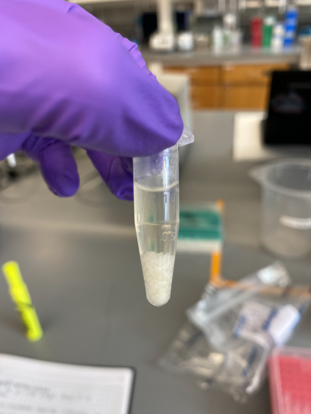
- The tubes were centrifuged again for 3 minutes at 16,000 rcf (g)
- A pellet was visible again in each tube, it looked smaller
- The supernatant was removed and discarded (hazardous)
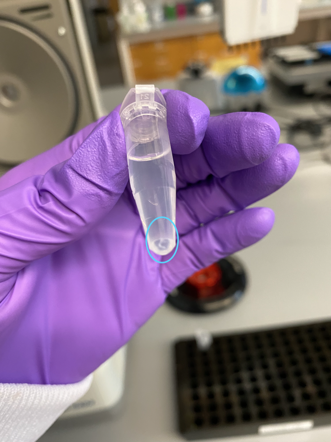
- Resuspended the pellet in 100ul high-salt TE buffer
- Diluted the Qiagen RNase A (100mg/mL) 1:100, 10ul RNase A to 990ul ultra pure water
- Added 5ul of diluted RNase A to each sample tube with the TE buffer (should be a final concentration close to 50ug/mL RNase A)
- Flicked to mix and spun down
- Incubated in the incubator genie (no rocking) for 15 minutes at 60 degrees C
- Afterwards tubes were brought to room temp by sitting for 5 minutes
- Added 900ul 80% ethanol to each tube and inverted to mix 10 times
- Centrifuged for 3 minutes at 16,000 rcf (g)
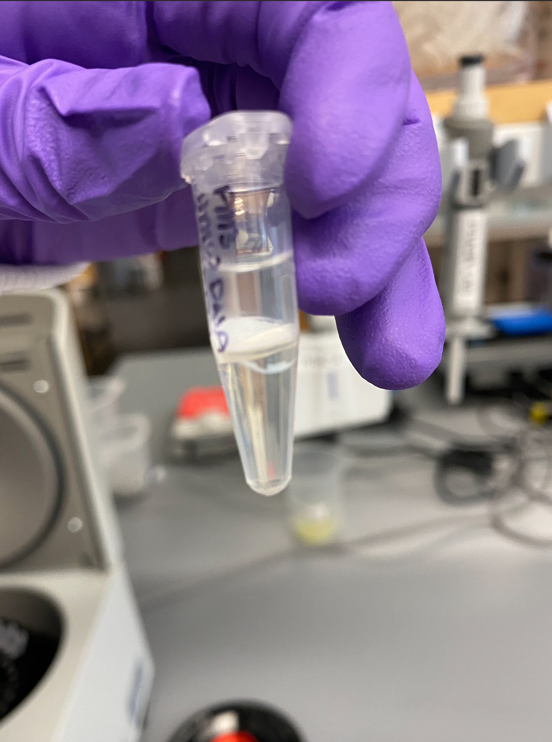
- There was no longer a visible pellet after this centrifugation but I removed the supernatant (hazardous) as if there was (avoided pipetting to the complete bottom of the tube)
- Added 1mL 80% ethanol to each tube and inverted to mix
- Centrifuged for 3 minutes at 16,000 rcf (g)
- There was a very small pellet visible in each tube again
- Discarded all of the supernatant (hazardous)
- Left tubes open in the hood for ~7 minutes to dry
- Added 50ul 10mM Tris HCl pH 8 to each tube
- Placed on rotating shaker room temp for 45 minutes to resuspend the DNA, 200rpm
QC
Qubit
- Broad Range DNA Qubit of the top and bottom of each tube, sometimes HMW DNA can clump in one part of the tube, that was not the case with these samples though
- Followed protocol
| Sample | Standard 1 (RFU) | Standard 2 (RFU) | Average DNA two readings (ng/ul) | Average Top and Bottom DNA (ng/ul) |
|---|---|---|---|---|
| PM1 T | 188 | 20031 | 32.1 | - |
| PM1 B | - | - | 32 | 32.05 |
| PM2 T | - | - | 24.6 | - |
| PM2 B | - | - | 24.8 | 24.7 |
| PM3 T | - | - | 14.3 | - |
| PM3 B | - | - | 14.8 | 14.5 |
| PM4 T | - | - | 32 | - |
| PM4 B | - | - | 32.3 | 32.15 |
| PM5 T | - | - | 24.4 | - |
| PM5 B | - | - | 25.5 | 24.9 |
| PM6 T | - | - | 15.7 | - |
| PM6 B | - | - | 15.3 | 15.5 |
TapeStation
- Followed protocol using the Genomic DNA screentape and reagents
- Results link
- The tapestation didn’t recognize the first band in the ladder, which is at 48,000bp, so the sizing is off. We are likely using a ladder that is just too old because it has happened before. I can run these samples again using a different ladder, but my expectation is the smear pattern will be the same. There is a lot of low bp DNA and not that big of a peak of HMW DNA. These are not good samples to send for PacBio sequencing