PCR of p47 (DiNV) and COI (control) on Gel Extraction Products from Last Two - Extractions to Determine if Virus DNA is in the HMW Fractions
20211117 COI PCR
- First needed to make personal diluted stocks of the COI primers
| Stock number | primer number | ul 100uM primer | ul molecular grade water for 10uM |
|---|---|---|---|
| 4 | 1490 | 10 | 90 |
| 5 | 2198 | 10 | 90 |
- Planned to run all samples from both extractions that had measurable DNA, plus a negative and positive control, equalling 13 samples, plus 2 for error. So I made enough mix for “15”
- The positive control is a from Kent and is a positive control for p47 (virus)
- All samples (except for the positive control) have low DNA (less than 2ng/ul), so I decided the DNA input would be 6ul for all samples (instead of the usual 1ul, however that’s often 10ng total DNA)
- All samples were kept on ice, along with primers, and enzymes
- Made master mix on ice:
- 1ul 10X NEB taq buffer * 15 = 15ul
- 1ul 2mM dNTPs * 15 = 15ul
- 0.25ul 10uM 1490 primer * 15 = 3.75ul
- 0.25ul 10uM 2198 primer * 15 = 3.75ul
- 0.1ul NEB Taq * 15ul = 1.5ul
- 1.4ul molecular grade water * 15 = 21ul
- Master mix was vortexed, spun down, and kept on ice
- 4ul of master mix was added to each tube of 13 strip tubes (kept on ice)
- 6ul of DNA from samples was added to their respective reaction tube, 6ul of molecular grade water was added to the negative control, and 5ul of water and 1ul of DNA was added to the positive control. (see table below)
| Tube # | Sample | ul DNA |
|---|---|---|
| 1 | 120A | 6 |
| 2 | 120B | 6 |
| 3 | 120A UPPER | 6 |
| 4 | 120A LOWER | 6 |
| 5 | 120B UPPER | 6 |
| 6 | 120B LOWER | 6 |
| 7 | 120A WELL | 6 |
| 8 | 120B WELL | 6 |
| 9 | 120 | 6 |
| 10 | 120 Top | 6 |
| 11 | 120 Bottom | 6 |
| 12 | Negative control | 6ul molecular grade water |
| 13 | Positive control | 5ul molecular grade water, 1ul DNA |
- Strip tubes were vortexed and spun down until no bubbles
- Tubes were placed in the PCR machine, in a new program COI, which is based off the one in Tom’s folder but with 30 cycles
- 94 degrees C 2 min
- 94 degrees C 20 sec
- 52 degrees C 20 sec
- 72 degrees C 1 min 30 sec
- 72 degrees C 5 minutes
- 12 degrees C hold
- bold fields are cycled 30 times
- Run time was 1 hour and 45 minutes
- Tubes were put in the 4 degree fridge overnight until gelling, tubes are labeled with primer name and date
20211117 p47 PCR
- Used the same samples, same order, same style mastermix for the p47 PCR
- The positive control is a from Kent and is a positive control for p47 (virus)
- All samples were kept on ice, along with primers, and enzymes
- Made master mix on ice:
- 1ul 10X NEB taq buffer * 15 = 15ul
- 1ul 2mM dNTPs * 15 = 15ul
- 0.25ul 10uM p47_F primer * 15 = 3.75ul
- 0.25ul 10uM p47_R primer * 15 = 3.75ul
- 0.1ul NEB Taq * 15ul = 1.5ul
- 1.4ul molecular grade water * 15 = 21ul
- Master mix was vortexed, spun down, and kept on ice
- 4ul of master mix was added to each tube of 13 strip tubes (kept on ice)
- 6ul of DNA from samples was added to their respective reaction tube, 6ul of molecular grade water was added to the negative control, and 5ul of water and 1ul of DNA was added to the positive control. (see table below)
| Tube # | Sample | ul DNA |
|---|---|---|
| 1 | 120A | 6 |
| 2 | 120B | 6 |
| 3 | 120A UPPER | 6 |
| 4 | 120A LOWER | 6 |
| 5 | 120B UPPER | 6 |
| 6 | 120B LOWER | 6 |
| 7 | 120A WELL | 6 |
| 8 | 120B WELL | 6 |
| 9 | 120 | 6 |
| 10 | 120 Top | 6 |
| 11 | 120 Bottom | 6 |
| 12 | Negative control | 6ul molecular grade water |
| 13 | Positive control | 5ul molecular grade water, 1ul DNA |
- Strip tubes were vortexed and spun down until no bubbles
- Tubes were placed in the PCR machine p47 program in the Maggie folder:
- 94 degrees C 5 minutes
- 94 degrees C 45 seconds
- 55 degrees C 1 minute
- 68 degrees C 1.5 minutes
- 68 degrees C 5 minutes
- Hold at 12 degrees C
- bold text is cycled through 30 times
- Run time was 1 hour and 20 minutes
- Tubes were put in the 4 degree fridge overnight until gelling, tubes are labeled with primer name and date
20211118 Gel of PCR Products
- 1% gel: 0.9g agarose and 90mL 1X TAE, microwaved ~3 min
- Used the gel tray with the red tape lines, took about 60-70mL of gel
- Loaded a 1kb plus ladder in the first well and in the well between the COI and the p47 samples
- Loaded all the COI samples, then the p47 samples
- Ran the gel for 1 hour at 90V
- Put the gel in the EtBr stain for 45 min (there was a little tear in the gel when transferring it to the stain, but it should still be able to be imaged)
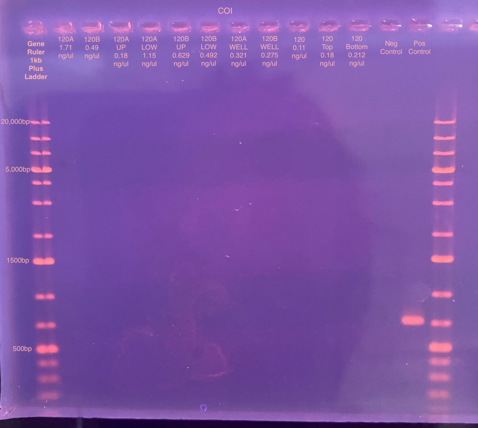
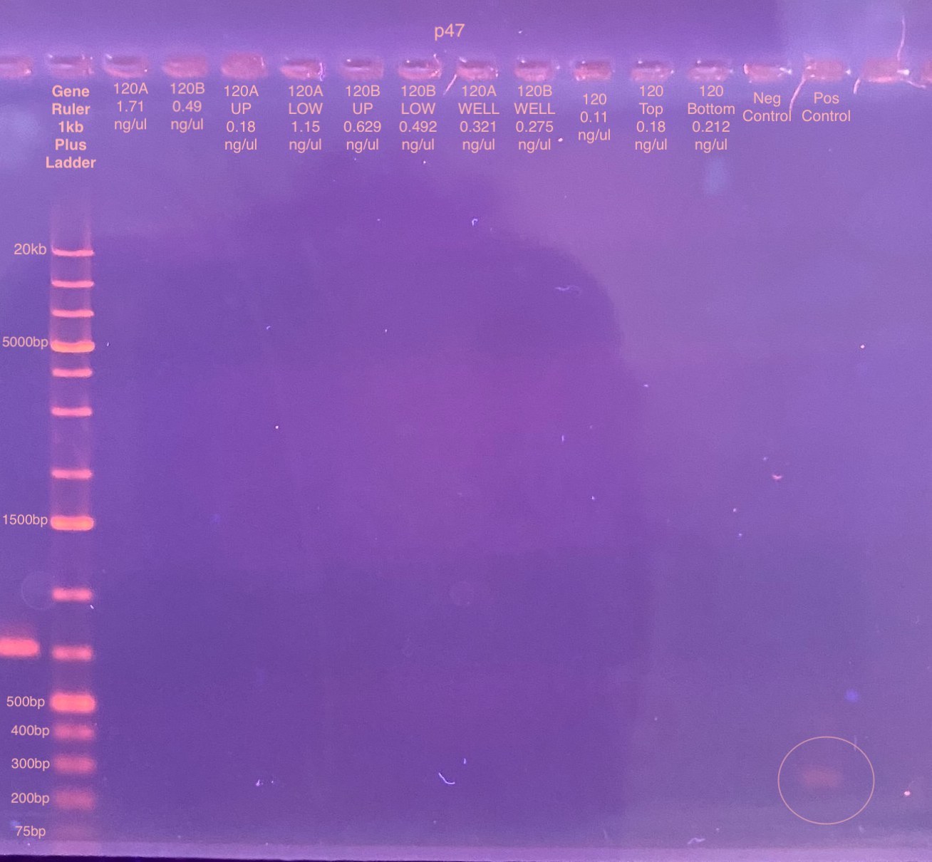
Looks like none of my extraction PCRs worked. I’m glad I have the positive control sample in there, I was not sure if would amplify for COI (I don’t know what it is), but clearly it did. I probably should have used one of my HMW extracted DNA samples as another positive control for this. I am pretty certain that the faint band in the p47 half of the gel is the amplification for the positive control…
I am not sure why there isn’t any amplifications in my extractions. Potentially there isn’t actually and DNA and the Qubit was wrong. Or there was a lot more DNA, Qubit was wrong again, and so I overloaded the reaction and nothing amplified. Or the elution liquid or the solution is not the right conditions for PCR, maybe salt or ethanol came out in the elution. Or there is left-over agarose in the elution preventing amplification.