Testing Cell Transfection Reagents and pAc5 GFP Expression Plasmid on Primary innubila Cells with S2 Cells as a Control
- This time I decided to do a more controlled experiment where I used S2 cells as a positive control, and I decided to make the primary cells the day before the transfection so they would be the most alive/most likely to be dividing
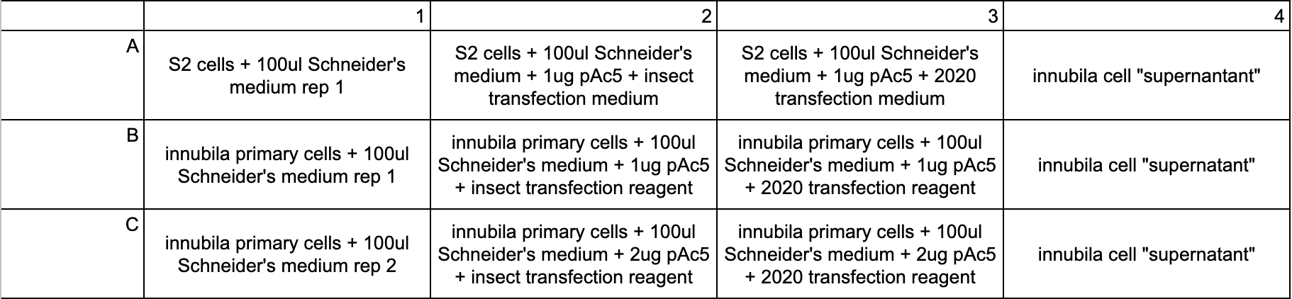
- On 10/25 I plated S2 cells in wells A1-A3, and plated fresh primaries in wells B1-3 and C1-3. Primary “supernatant” is in column 4 in the plate, but they were not used for the experiment
- The only controls used were cells without plasmid or reagent
- I tried both a 1:1 ratio of plasmid DNA to reagent, and 2:1 plasmid DNA to reagent. The plasmid DNA is at 1ug/ul, so adding 2ul instead of 1ul doubles the amount of plasmid added
- Because of the plasmid DNA ratio increase, technically there are no replicates for the experimental conditions, the only replicate is the plain primary cells
- Plate layout:
- All of this was done at room temperature in the cell culture hood. Transfection reagents were thawed at room temp, vortexed, and spun down before use
- Reagent tubes were made for each experimental condition:
- Tube 1:
- 300ul Schneider’s medium
- Tube 2:
- 200ul Schneider’s medium
- 2ul pAc5 plasmid
- 4ul insect transfection reagent
- Tube 3:
- 100ul Schneider’s medium
- 2ul pAc5 plasmid
- 2ul insect transfection reagent
- Tube 4:
- 200ul Schneider’s medium
- 2ul pAc5 plasmid
- 6ul 2020 reagent
- Tube 5:
- 200ul Schneider’s medium
- 2ul pAc5 plasmid
- 3ul 2020 reagent
- Tube 1:
- All tubes were pipette mixed and incubated in the hood for 30 minutes
- Then the reagent mixes were dropped into the wells:
- A1: 100ul tube 1
- A2: 103ul tube 2
- A3: 104ul tube 4
- B1: 100ul tube 1
- B2: 103ul tube 2
- B3: 104ul tube 4
- C1: 100ul tube 1
- C2: 104ul tube 3
- C3: 105ul tube 5
- The plate was gently rocked back and forth and put in the 23 degree incubator for 24 hours, then checked. The plate was always kept in the incubator between checks
20221027 Checking transfections
- S2 cells with the transfection reagents had a few GFP cells after 24 hours, and the amount grew over the days of checking them
- No innubila experimental wells showed any evidence of GFP after 24 or more hours
- Again there was a lot of autofluorescence in the primary cells so it’s very hard to tell if there is GFP or not, but it really looks the same as the controls, and I didn’t see any of the neon green indicative of GFP
- I imaged the plates in a much more comprehensive manner this time, and I also included bright field images, seen here